Newcastle Disease (NDV) current situation in Morocco


Detection of Inclusion Body Hepatitis Virus in Broiler Chickens Using Genetic Analysis Techniques
Dr. Hisham Alma’aita, PhD in Poultry Diseases
Manager of Poultry Health, National Poultry Company
Abstract
Inclusion Body Hepatitis (IBH) is an economically significant viral disease in poultry, caused by Fowl
Adenoviruses (FAdVs) belonging to the genus Aviadenovirus, which comprises five species (FAdV-A to
FAdV-E) with multiple serotypes. Among these, species FAdV-E—which includes 12 serotypes—is most
associated with IBH, Hydropericardium Syndrome (HPS), and immunosuppression in broiler chickens.
Recently, broiler farms reported increased mortality ranging from 10–20%, accompanied by general
weakness, reduced feed intake, stunting, and apathy. Post-mortem examination revealed hepatomegaly, pale
or yellow discoloration of the liver with friable consistency, and the presence of hemorrhagic or necrotic
foci on the hepatic surface, in addition to clear hydropericardium.
A total of ten liver samples and four pericardial fluid samples were collected from suspected broilers and
preserved on FTA cards for laboratory testing.
Polymerase chain reaction (PCR) assays targeting the hexon gene detected FAdV in four liver samples and
all four pericardial fluid samples. Partial sequencing of the ~800 bp hexon gene fragment, followed by
phylogenetic analysis and serotype classification (species A–E; serotypes 1–7, 8a, 8b, 9–11), revealed that
all PCR-positive isolates clustered within serotype 8b of species FAdV-E (designated FAdV-E/8b)
The findings confirm that FAdV serotype 8b strains are not only primary etiological agents of IBH and
HPS—with associated mortality reaching up to 20%—but are also capable of inducing immunosuppression,
increasing susceptibility to secondary infections and impairing vaccine responses.
The study recommends implementing strict preventive strategies against FAdV infection, particularly
through vaccination of breeder flocks using homologous or genetically matched vaccines. This approach
aims to ensure the transfer of robust serotype-specific maternal immunity to broilers, thereby reducing
economic losses in poultry production systems.
Detection of Inclusion Body Hepatitis Virus in Broiler Chickens
1
Materials and Methods
Broiler Chickens
Broiler chickens exhibiting clinical signs including general weakness, lethargy, reduced feed intake, growth
retardation, and increased mortality rates ranging between 10–20% were examined and sampled for this
study.
Samples
A total of ten liver samples and four pericardial fluid samples from birds showing hydropericardium were
collected. All samples were preserved on FTA cards and submitted to the laboratory for the detection of
Fowl Adenovirus (FAdV).
DNA Extraction from FTA Cards
Small sections were excised from each FTA card sample and transferred into sterile microcentrifuge tubes.
Subsequently, 500 µL of TE buffer was added to each tube, followed by vortexing. The tubes were incubated
at room temperature for 24 hours to allow release of nucleic acids from the card matrix. After incubation,
100 µL of the recovered sample was transferred into 300 µL of TRIzol reagent, mixed thoroughly, and
incubated for 5 minutes prior to downstream processing.
Real-Time PCR Amplification
The hexon gene of FAdV was amplified using conventional PCR with in-house–designed primers. The
expected amplicon size was approximately 800 base pairs (bp). Primer nucleotide sequences were as
follows:
• adeno-F: 5′-ACATGGGAGCGACCTACTTCGACA-3′
• adeno-R: 5′-TCGGCGAGCATGTACTGGTAAC-3′
Genetic Analysis of the Hexon Gene
Liver and pericardial fluid samples that tested positive by PCR were selected for partial sequencing of the
hexon gene (~800 bp). Phylogenetic analysis and serotype classification (species A–E; serotypes 1–7, 8a,
8b, 9–11) were performed. Forward and reverse primers were used in two independent reactions based on
the purified nucleic acid extract.
Detection of Inclusion Body Hepatitis Virus in Broiler Chickens
2
Results
Clinical Signs
Affected broiler flocks exhibited prominent clinical signs including lethargy, huddling behavior, ruffled
feathers, reduced feed intake, and yellow mucoid diarrhea. A marked decline in growth performance was
observed, leading to deterioration in feed conversion ratio.
In more advanced stages of infection, chicks displayed severe depression, reduced mobility, and a tendency
to cluster in the corners of the house. Mortality ranged between 10–20%, with peak mortality occurring on
days 3–4 following the onset of clinical signs (Figure 1).
Post-mortem Lesions
Post-mortem examination revealed characteristic lesions in vital organs including the liver, heart, and
kidneys. The liver appeared markedly enlarged, pale to yellowish, friable, and showed multifocal necrotic
and pinpoint hemorrhagic lesions.
Hydropericardium was consistently observed, characterized by an accumulation of clear to straw-colored
serous fluid within the pericardial sac, sometimes reaching volumes up to 10 mL, giving the heart a
balloon-like appearance.
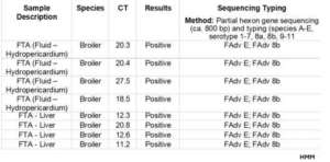
Real-time PCR Detection
PCR assays targeting the hexon gene detected FAdV in all four pericardial fluid samples and four out of ten
liver samples. Cycle threshold (Ct) values ranged between 18.5–27.5 for pericardial fluid samples and 11.2
20.8 for liver samples, indicating variable viral loads among the tested tissues (Table 1).
Genetic Analysis of the Hexon Gene
Partial sequencing of the hexon gene from all PCR-positive samples (four liver samples and four pericardial
fluid samples) confirmed the presence of Fowl Adenovirus serotype 8b, belonging to species FAdV-E.
Phylogenetic analysis demonstrated that all isolates clustered tightly with reference strains of FAdV-E/8b,
as shown in Table 1.
Detection of Inclusion Body Hepatitis Virus in Broiler Chickens
3
Figure 1.
Clinical signs observed in broiler flocks affected by Inclusion Body Hepatitis (IBH), including lethargy,
huddling behavior, ruffled feathers, reduced feed intake, and increased mortality.
Figure 2.
Gross pathological lesions of the liver in broiler chickens infected with Inclusion Body Hepatitis (IBH),
showing hepatomegaly, pale or yellow discoloration, friable texture, and multifocal necrotic and
hemorrhagic lesions.
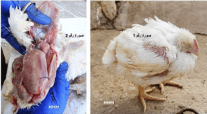
Table 1.
Real-time PCR results and partial hexon gene sequencing data for pericardial fluid and liver samples from
broiler chickens, confirming the detection and molecular characterization of Fowl Adenovirus (FAdV)
serotype 8b (FAdV-E/8b).
Recommendations
1. Identification of the circulating FAdV strain is essential for understanding the epidemiology of the
disease.
2. The detection of FAdV-8b—a serotype strongly associated with Inclusion Body Hepatitis (IBH)
outbreaks in broilers—provides critical insights that support:
o Tracing the source of infection and determining locally or regionally prevalent strains.
o Developing more precise vaccination programs tailored to the dominant field strains.
o Strengthening biosecurity and disease-control strategies to minimize economic losses in
poultry production.
o Supporting epidemiological studies that correlate detected serotypes with disease severity
and flock mortality patterns.
3. Effective control of IBH requires strict biosecurity measures, optimal flock management,
prevention and control of immunosuppressive diseases, and the strategic use of vaccination in
endemic regions.
4. In areas with widespread IBH occurrence, the use of inactivated (killed) adenovirus vaccines
containing the circulating strain—specifically FAdV-E/8b—is recommended for breeder flocks.
This ensures the transfer of high levels of serotype-specific maternal antibodies to broiler chicks,
providing early protection against IBH and reducing associated mortality.
References
1. Adel, A., Mohamed, A. E., Samir, M., Hagag, N. M., Erfan, A., Said, M., & Shahien, M. A. (2021).
Epidemiological and molecular analysis of circulating fowl adenoviruses and emergence of
serotypes 1, 3, and 8b in Egypt. Heliyon, 7(12), e08543.
2. Al-Hallaq, E., & Bakri, H. (2020). Molecular detection and characterization of Fowl Adenovirus in
broiler chickens in Jordan. Avian Pathology, 49(4), 1–9.
3. Bakri, H., Al-Hallaq, E., et al. (2021). Pathological investigation on Fowl Adenovirus infection in
the Middle East (2019–2021). Vaxxinova International BV (Technical Report).
4. Hussein, M., et al. (2019). Prevalence of Fowl Adenovirus in commercial broiler farms in Jordan.
Jordan Journal of Agricultural Sciences, 15(3), 1–12.